We have designed a novel positron emission tomography radiotracer, '18F"PKM2-23, for the noninvasive imaging of tumor pyruvate kinase M2 (PKM2) with positron emission tomography (PET). Aberrant tumor glycolysis is partly mediated by PKM2, which controls the balance between energy production and the synthesis of metabolic precursors.
Stanford researchers at the Gambhir Lab have designed a new PET radiotracer, [
18 F]PKM2-23, for the non-invasive imaging of tumor pyruvate kinase M2 (PKM2) with positron emission tomography (PET). The increased glucose utilization of tumors in comparison to normal tissue has previously been exploited clinically with [
18 F]FDG PET. This new radiotracer can to detect malignancies where FDG (fluorodeoxyglucose) fails, for example, in the brain where FDG background is high. The team demonstrated the radiolabeling of PKM2-23 with fluorine-18 and reported its ability to retain specificity for PKM2. Radiolabeling of PKM2-23 with fluorine-18 is advantageous for clinical translation due to its very long half-life of 110min. The synthesis method also has economic and temporal advantages over current methods. Thus, healthcare providers could effectively and noninvasively detect, stage, and monitor disease progression in brain tumors using this new radiotracer.Figure
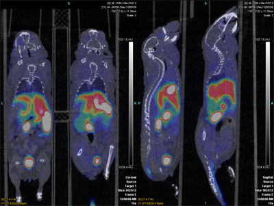
Figure 1 -60 min summed dynamic image of [
18 F]DASA-23 in nude mice Dynamic PET imaging with [
18 F]DASA-23 (Figure 1) revealed rapid liver and kidney uptake and clearance through both renal and hepatobiliary routes. The initial high, delivery of [
18 F]DASA-23 in the brain peaked 30s after injection (5.2 ± 0.5% ID/g), this was followed by rapid clearance, reaching low steady-state levels (1.8 ± 0.1% ID/g) by 10min after injection. These properties are ideal for brain imaging of malignancies with [
18 F]DASA-23. [
18 F]DASA-23 has shown the ability to delineate tumor margins in orthotopically growing models of human glioblastoma. U87 cells were implanted intracranially into nude mice and imaged with [
18 F]DASA-23 18days post implantation. [
18 F]DASA-23 clearly allowed for the detection of intracerebral tumors, as highlighted in the fused PET/CT/MRI image in Figure 2.
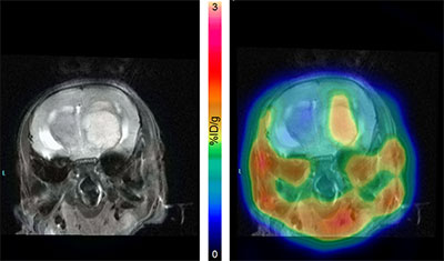
Figure 2. MRI (left) and fused PET/CT/MRI (right) images of [
18 F]DASA-23 in orthotopic human U87 tumors.
 May 9, 2017


 Premium